
Peripheral angiography is a minimally invasive imaging procedure used to examine blood flow through the arteries and veins outside the heart, particularly those supplying the legs. It can help identify narrowing, blockages, or other abnormalities affecting circulation.
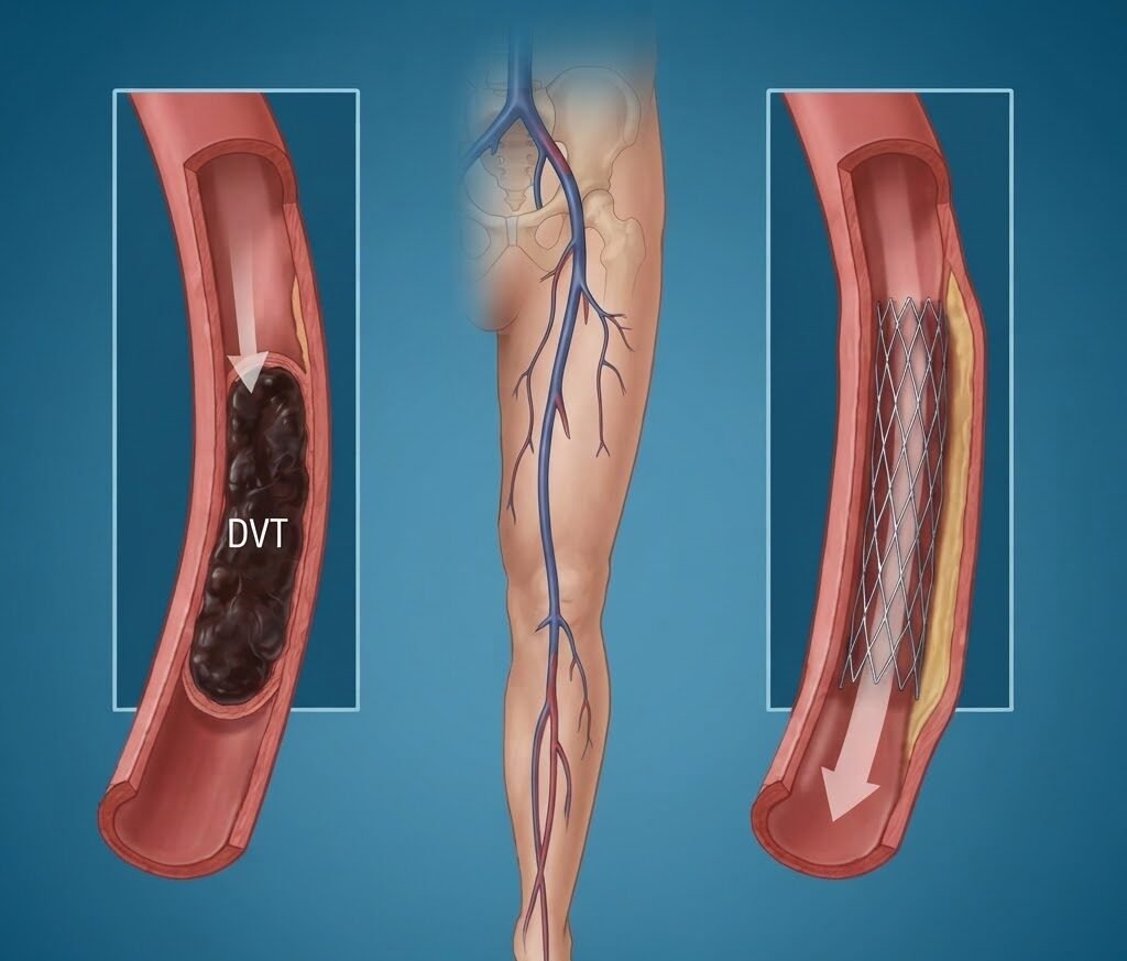
DVT (Deep Vein Thrombosis) is a condition in which a blood clot develops in a deep vein, most commonly in the leg. Depending on the location, severity, symptoms, and risk of complications, appropriate medical or interventional treatment may be recommended.
At Dr. Manish Juneja’s cardiac care practice in Nagpur, patients with suspected vascular problems can undergo detailed evaluation to determine the most appropriate treatment approach.
What Is Peripheral Angiography?
Peripheral angiography uses a thin catheter and contrast dye along with X-ray imaging to visualize blood vessels in areas such as the legs. It helps doctors identify reduced blood flow caused by narrowed or blocked arteries.
Depending on the findings, treatment options may include medicines, angioplasty, stenting, or other vascular interventions.
What Is DVT?
Deep Vein Thrombosis (DVT) occurs when a blood clot forms in a deep vein, usually in the lower leg or thigh. Common symptoms may include:
- Swelling in one leg
- Leg pain or tenderness
- Warmth around the affected area
- Red or discolored skin
DVT requires timely medical evaluation because a clot can sometimes travel to the lungs and cause a potentially serious condition called pulmonary embolism.
When May Peripheral Intervention Be Recommended?
A vascular intervention may be considered when there is significant reduction in blood flow or a blockage that requires treatment. Depending on the condition, the doctor may recommend:
- Peripheral angioplasty
- Stent placement
- Catheter-based clot treatment in selected cases
- Medication and monitoring
- Further vascular evaluation
The treatment is selected according to the patient’s symptoms, imaging findings, and overall health.
How Is Peripheral Angiography Performed?
1. Patient Evaluation
The doctor reviews symptoms, medical history, medications, and relevant blood or imaging tests before the procedure.
2. Catheter Insertion
A thin catheter is introduced through an appropriate blood vessel, commonly through the groin or another suitable access point.
3. Contrast Injection
Contrast dye is administered through the catheter so that the blood vessels can be clearly visualized.
4. Imaging
X-ray imaging is used to identify narrowing, blockage, or abnormal blood flow in the peripheral vessels.
5. Treatment if Required
If a significant blockage is identified, angioplasty, stenting, or another appropriate intervention may be considered depending on the patient’s condition.
Benefits of Peripheral Angiography & Interventions
- Helps identify vascular blockages and narrowing
- Provides detailed information about blood circulation
- Supports accurate treatment planning
- May help restore blood flow in selected patients
- Allows minimally invasive treatment when appropriate
- Helps evaluate complex peripheral vascular conditions.
Risks and Precautions
Peripheral angiography and vascular interventions are generally performed under medical supervision, but possible risks can include bleeding or bruising at the catheter site, blood vessel injury, infection, contrast-related reactions, or other procedure-related complications.
Patients should inform the doctor about kidney problems, diabetes, allergies, blood-thinning medicines, and previous reactions to contrast dye before the procedure.
Precautions After Peripheral Angiography & Intervention
1. Take Medicines as Prescribed
Take all prescribed medicines exactly as advised. Do not stop blood-thinning or other medications without consulting your doctor.
2. Care for the Catheter Site
Keep the access area clean and monitor it for bleeding, swelling, increasing pain, or unusual discharge.
3. Avoid Heavy Activity Initially
Avoid strenuous exercise and heavy lifting for the period recommended by your doctor, allowing the access site and body to recover.
4. Attend Follow-Up Appointments
If your doctor permits, adequate fluid intake may help your body clear the contrast material used during the procedure.
5. Monitor Your Leg Symptoms
Pay attention to increasing swelling, pain, color changes, numbness, or unusual coldness in the affected limb and report concerning symptoms promptly.
6. Attend Follow-Up Appointments
Follow-up visits allow the doctor to review your recovery, assess circulation, and modify treatment if necessary.